Medical image analysis method and program
No need to prepare mask images, and be capable of analyzing the tumor with surrounding tissues
Overview
Radiomics, a technique for calculating quantitative features, is used to diagnose tumors in medical images. However, this technique requires preparation of a mask image from which the outline of the affected area is extracted, which places a heavy burden on physicians. In addition, since the target of the calculation is limited to the area surrounded by the mask image, the relationship between the tumor and the surrounding tissue cannot be evaluated.
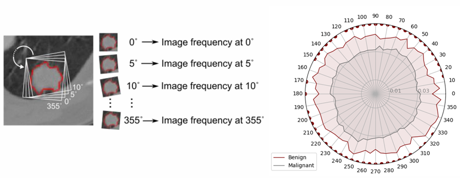
This technology relates to a tumor image diagnosis method and program that calculates the image frequency included in the transition of the correlation coefficient by enclosing the area including the tissue surrounding the tumor in a square and performing Fourier analysis on the correlation coefficient of the area.
Features:
1. No mask image required, just specify the user interested area by drawing a square
2. The relationship between tumor and surrounding tissue can be analyzed
3. Fast calculation (a few seconds per case)
360° analyzing pulmonary nodule evaluation image (left) and benign/malignant results (right)
Product Application
・Medical image analysis software (e.g. CT)
Expectations of companies
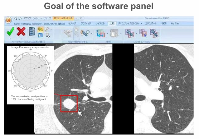
If you are interested in developing image viewer software like the one shown in the upper right, please contact us.
IP Data
IP No. : JP2024-208613
Inventor : USUZAKI Takuma
keyword : Medical image analysis software, CT, Tumor diagnosis